
Медицинская визуализация
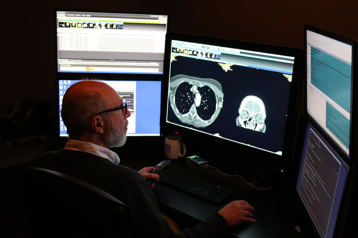
Отделение медицинской визуализации использует передовые технологии для обеспечения точной диагностики и планирования лечения. Иногда ключ к выздоровлению кроется в том, что мы не можем увидеть невооруженным глазом. Наша команда специалистов по медицинской визуализации использует современные технологии, такие как рентген, МРТ, КТ и УЗИ, чтобы помочь вашим лечащим врачам понять, что происходит внутри вашего организма. Но дело не только в оборудовании — дело в людях, которым небезразлично ваше здоровье. Мы понимаем, что прохождение обследований может вызывать беспокойство, поэтому уделяем время тому, чтобы объяснить вам процесс, ответить на ваши вопросы и обеспечить вам комфорт. Наши радиологи и технические специалисты работают с точностью и состраданием, предоставляя результаты, которые служат основой для постановки диагноза и назначения лечения. Независимо от того, исследуете ли вы новый симптом или наблюдаете за хроническим заболеванием, мы всегда готовы поддержать вас, предоставляя четкую информацию и проявляя доброжелательность.
Мы можем помочь вам в следующих вопросах:
- Современные методы визуализации: КТ, МРТ, ПЭТ/КТ
- Визуализация молочной железы:маммография, комплексное обследование молочной железы
- Здоровье костей: DXA(оценка плотности костной ткани)
- Специализированная диагностическая визуализация:ядерная медицина
- Рутинная диагностическая визуализация:рентгенография, ультразвуковое исследование
Frequently Asked Questions: CT Scan
A computed tomography (CT) scan is a noninvasive medical test that helps physicians diagnose and treat medical conditions. CT combines x-ray equipment with computers to produce multiple cross-sectional images of the body. These images can then be examined on a computer monitor.
The CT exam lasts about 10-15 minutes, unless you are drinking oral contrast or are having a specialized exam such as a biopsy or other procedure. Drinking oral contrast may add 30 minutes to two hours. Total time you will be at the healthcare facility is 30 minutes to 3 hours. This information will be available to you when you schedule your appointment. You should wear comfortable, loose-fitting clothing for your exam. You may be given a gown to wear during the procedure. Metal objects in the area being scanned, including jewelry, eyeglasses, dentures and hairpins, may affect the CT images and should be left at home or removed prior to your exam. You may also be asked to remove hearing aids and removable dental work. You may be asked to remove any piercings, if possible.
В зависимости от типа обследования вам может быть рекомендовано воздержаться от еды и питья за несколько часов до его начала. Возможно, вам установят капельницу, и в ходе обследования будет использоваться внутривенное контрастное вещество, или «краситель». Вам следует сообщить врачу обо всех лекарствах, которые вы принимаете, а также о наличии аллергии. Если у вас известна аллергия на внутривенное контрастное вещество, или «краситель», врач может назначить лекарства (обычно стероиды) для снижения риска аллергической реакции. Эти лекарства, как правило, необходимо принять за 12 часов до введения внутривенного контрастного вещества.
Inform your doctor of any recent illnesses or other medical conditions and whether you have a history of heart disease, asthma, diabetes, and kidney disease or thyroid problems. Any of these conditions may increase the risk of an unusual adverse effect to the contrast media. Women should always inform their physician and the CT technologist if there is any possibility that they may be pregnant.
В случае с детскими пациентами родителю может быть разрешено находиться в кабинете, но при этом он должен будет надеть свинцовый фартук или сидеть за свинцовой перегородкой, чтобы свести к минимуму облучение.
Frequently Asked Questions: Fluoroscopy
Флюороскопия — это особый вид рентгенографии. С помощью флюороскопии рентгеновские снимки можно просматривать как рентгеновский фильм, и радиолог может наблюдать за движением внутренних органов, например толстой кишки. Флюороскопия также помогает радиологу понять, как некоторые органы, например желудок, опорожняются от принятой пищи, и определить, нормально ли функционирует желудок. Рентгенологи и рентгенотехники проходят специальную подготовку, чтобы использовать минимально необходимое количество рентгеновского излучения для получения диагностического изображения. Эти специалисты знают, как безопасно проводить обследование как взрослых, так и детей.
Depending on the exam ordered, your physician may ask you not to eat or drink for several hours beforehand or provide specific preparation instructions. Some exams also require blood work to be completed before the imaging study. During your exam, you may be asked to change into a gown and remove jewelry, eyeglasses, dental appliances, or any metal objects or clothing that could interfere with the images. If there is any possibility you are pregnant, please inform your physician or technologist before the exam. Most imaging tests are avoided during pregnancy to limit radiation exposure. If an X‑ray is necessary, special precautions will be taken to protect you and your baby. Depending on the area being examined, images may be taken while you are standing or lying on an X‑ray table. Multiple images from different angles may be needed. Exam times typically range from 30 minutes to 2 hours.
For Barium Studies: If you do not have a bowel movement for more than 2 days after your exam, or you cannot pass gas through your rectum.
For Contrast Studies, complications are rare. If you were given contrast, call your doctor if any of the following occurs after the exam:
- Крапивница
- Зуд
- Тошнота
- Отек и зуд в глазах или на губах
- Сдавленность в горле
- Затрудненное дыхание
When the examination is complete, you may be asked to dress and wait while the images are reviewed. Depending on the exam type, you may be asked to drink plenty of fluids to flush the barium or contrast out of your system. If you had barium, your stools may appear white, as your body clears the barium from your system. A radiologist will review the images and send a report to your referring doctor. You will receive your results from the doctor who ordered the test.
Frequently Asked Questions: X-Ray
Рентгенография — это метод медицинской визуализации, позволяющий получать изображения внутренних органов и тканей организма, что помогает врачам ставить диагнозы. К распространенным видам рентгенографии относятся снимки грудной клетки, брюшной полости, позвоночника, черепа, околоносовых пазух и конечностей. В некоторых случаях для лучшей визуализации определенных органов или тканей используется контрастное вещество или барий; такие исследования проводятся с помощью флюороскопии. Радиологи и рентгенотехники проходят специальную подготовку, чтобы использовать минимально необходимое количество излучения для получения четких диагностических изображений и безопасно проводить обследования как взрослых, так и детей.
Most x-rays require no special preparation. You may be asked to remove some or all of your clothes and to wear radiology attire during the exam. You may also be asked to remove jewelry, removable dental appliances, eyeglasses and any metal objects or clothing that might interfere with the x-ray images. Women should always inform their physician and x-ray technologist if there is any possibility that they are pregnant. Many imaging tests are not performed during pregnancy so as not to expose the fetus to radiation. If an x-ray is necessary, precautions will be taken to minimize radiation exposure to the baby.
When the examination is complete, you may be asked to dress and wait while the images are reviewed. A radiologist will review the images and send a report to your referring doctor. You will receive your results from the doctor who ordered the test.
Depending on the area of interest, you may be placed on the x-ray table, or the exam may be done standing. Various x-rays may be taken at different angles to best visualize the area. The duration of the exam ranges from 15 to 30 minutes, depending on the type of x-ray or number of images ordered by your doctor. Barium and contrast studies may take from 30 minutes to 2 hours.
Frequently Asked Questions: MRI
Магнитно-резонансная томография (МРТ) использует мощное магнитное поле, радиоволны и компьютер для получения подробных изображений внутренних структур организма. Она применяется для диагностики различных состояний, включая опухоли и заболевания печени, сердца и кишечника. МРТ является неинвазивным методом и не предполагает использования ионизирующего излучения.
MRI exams typically take 45 minutes to one hour per body part. If contrast is used, the exam may take an additional 15–20 minutes. Plan to spend one to two hours total at the facility. Little preparation is usually required. You may take your regular medications unless instructed otherwise. If fasting or other dietary restrictions are needed, you will be notified when scheduling. Please arrive 15–30 minutes before your scheduled exam time, as instructed during scheduling. You will change into radiology attire for your exam. Jewelry, hair accessories, and other metal items are not permitted. A locker will be provided for your belongings. If you have any implanted medical devices—such as electronic implants, vascular or cardiac stents, or other surgical implants—please bring your device card or related documentation. This information is required to confirm that the device is safe for MRI.
If you had IV contrast material, the technologist would remove the IV and place a bandage on the IV site. Drink plenty of fluids to help flush all contrast media out of your system. If you receive sedation, someone must drive you home. Your images will be read by a radiologist, and a report will be sent to your doctor. Your doctor will get results of your exam within a few days.
An MRI machine is a large, cylinder‑shaped magnet that is open at both ends. You will lie on a moveable table that slides into the center of the magnet for imaging. Before your exam, you will complete an MRI safety screening form. These questions may be asked when scheduling and will be reviewed again by the MRI technologist before your scan. If IV contrast is part of your exam, the technologist will place an IV beforehand. You will need to lie very still during the scan, as movement can blur the images and extend the exam time. Depending on the area being examined, you may be asked to hold your breath for up to 30 seconds at a time. The MRI suite is well lit and includes a fan for comfort. You can communicate with the technologist at any time using a two‑way intercom, and you’ll be given an alarm button to alert staff if you feel uncomfortable. During the exam, you will hear loud, intermittent knocking sounds. Earplugs or headphones will be provided, and you may choose to listen to music. If you experience claustrophobia, talk with your provider in advance. They may prescribe medication to help you relax, and you’ll need someone to drive you home if medication is used.
Осложнения возникают крайне редко (менее 1 %).Если вам ввели контрастное вещество, обратитесь к врачу, если после обследования у вас возникнет какой-либо из следующих симптомов:
- Крапивница
- Зуд
- Тошнота
- Отек и зуд в глазах или на губах
- Сдавленность в горле
- Затрудненное дыхание
Frequently Asked Questions: Ultrasound
Ультразвуковое исследование — это метод медицинской визуализации, при котором с помощью звуковых волн создаются изображения внутренних органов. Во время обследования на кожу наносят гель и прикладывают небольшой ручной прибор, называемый датчиком. Датчик посылает звуковые волны вглубь тела, а по возвращающимся эхо-сигналам на компьютере формируются изображения. При ультразвуковом исследовании не используется излучение. Поскольку изображения получаются в режиме реального времени, ультразвуковое исследование позволяет наблюдать движение внутренних органов, а также кровоток в кровеносных сосудах.
Please wear comfortable, loose‑fitting clothing to your ultrasound exam. You may be asked to remove clothing and jewelry in the area being examined and change into a gown. Preparation varies depending on the type of ultrasound. Some exams require you to avoid eating or drinking for up to 12 hours beforehand. Others may require you to drink several glasses of water before your appointment and avoid urinating, so your bladder is full. You will receive specific instructions when your exam is scheduled.
During the exam, a trained ultrasound technologist (sonographer) will apply warm gel to your skin and move a small handheld device called a transducer over the area being examined. The transducer sends sound waves into the body and captures the returning echoes to create images. Multiple images may be taken by adjusting the transducer, and in some cases a radiologist may obtain additional images. Most ultrasound exams take 30 minutes to one hour to complete.
When the examination is complete, you may be asked to dress and wait while the ultrasound images are reviewed. After an ultrasound examination, you should be able to resume your normal activities immediately. A radiologist who specializes in ultrasound will review the images and send a report to your referring doctor. You will receive your results from the doctor who ordered the test.
Сертификаты и аккредитация
Lorem ipsum dolor sit amet, consectetuer adipiscing elit, sed diam nonummy nibh euismod tincidunt.


Связанные места

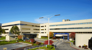
Больница Confluence Health | Центральный кампус
1201 S. Miller St.
, Венатчи, штат Вашингтон, 98801
Найдите подходящего специалиста
Ознакомьтесь с полным списком наших врачей, специалистов и медицинских бригад — воспользуйтесь фильтрами по услугам, языку, местоположению и страховке, чтобы найти подходящего для вас специалиста.