Головна Послуги Медична візуалізація

Медична візуалізація

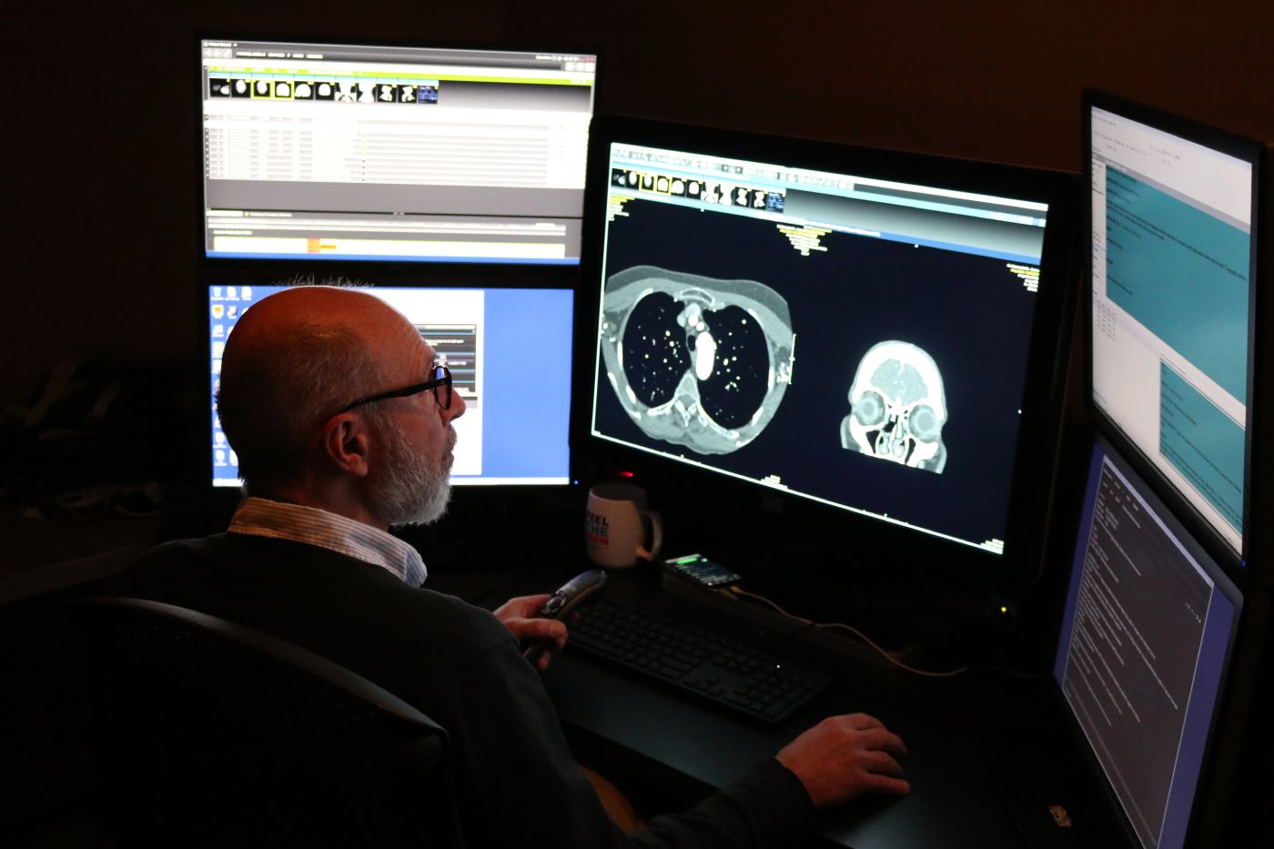
Медична візуалізація використовує передові технології для забезпечення точної діагностики та планування лікування. Іноді ключ до одужання криється в тому, чого ми не можемо побачити неозброєним оком. Наша команда фахівців з медичної візуалізації використовує сучасні технології, такі як рентген, МРТ, КТ та УЗД, щоб допомогти вашим лікарям зрозуміти, що відбувається всередині вашого тіла. Але справа не лише в апаратурі — а в людях, яким не байдуже. Ми розуміємо, що обстеження можуть викликати занепокоєння, тому ми приділяємо час, щоб пояснити процес, відповісти на ваші запитання та забезпечити ваш комфорт. Наші радіологи та технічні фахівці працюють з точністю та співчуттям, надаючи результати, які слугують основою для діагностики та лікування. Незалежно від того, чи ви досліджуєте новий симптом, чи контролюєте перебіг хронічного захворювання, ми завжди готові підтримати вас, надаючи чітку інформацію та проявляючи доброзичливість.

Ми можемо допомогти вам у таких питаннях:

  • Сучасні методи візуалізації: КТ, МРТ, ПЕТ/КТ
  • Візуалізація молочних залоз:мамографія, комплексний догляд за молочними залозами
  • Здоров’я кісток: DXA(дослідження щільності кісткової тканини)
  • Спеціальна діагностична візуалізація:ядерна медицина
  • Рутинна діагностична візуалізація:рентген, ультразвукове дослідження

Догляд починається тут


Frequently Asked Questions: CT Scan

A computed tomography (CT) scan is a noninvasive medical test that helps physicians diagnose and treat medical conditions. CT combines x-ray equipment with computers to produce multiple cross-sectional images of the body. These images can then be examined on a computer monitor.

The CT exam lasts about 10-15 minutes, unless you are drinking oral contrast or are having a specialized exam such as a biopsy or other procedure. Drinking oral contrast may add 30 minutes to two hours. Total time you will be at the healthcare facility is 30 minutes to 3 hours. This information will be available to you when you schedule your appointment. You should wear comfortable, loose-fitting clothing for your exam. You may be given a gown to wear during the procedure. Metal objects in the area being scanned, including jewelry, eyeglasses, dentures and hairpins, may affect the CT images and should be left at home or removed prior to your exam. You may also be asked to remove hearing aids and removable dental work. You may be asked to remove any piercings, if possible.

Залежно від типу обстеження вас можуть попросити нічого не їсти й не пити протягом кількох годин перед його початком. Вам можуть встановити крапельницю та під час обстеження використовувати внутрішньовенні контрастні речовини, або «барвник». Ви повинні повідомити лікаря про всі ліки, які ви приймаєте, а також про наявність будь-яких алергій. Якщо у вас є відома алергія на внутрішньовенні контрастні речовини, або «барвник», лікар може призначити ліки (зазвичай стероїди) для зменшення ризику алергічної реакції. Ці ліки, як правило, потрібно прийняти за 12 годин до введення внутрішньовенної контрастної речовини.

Inform your doctor of any recent illnesses or other medical conditions and whether you have a history of heart disease, asthma, diabetes, and kidney disease or thyroid problems. Any of these conditions may increase the risk of an unusual adverse effect to the contrast media. Women should always inform their physician and the CT technologist if there is any possibility that they may be pregnant.

У випадку з дітьми в палату може бути допущений один із батьків, але він повинен буде надіти свинцевий фартух або сидіти за свинцевою перегородкою, щоб мінімізувати опромінення.

Frequently Asked Questions: Fluoroscopy

Флюороскопія — це особливий вид рентгенографії. За допомогою флюороскопії рентгенівські знімки можна переглядати як рентгенівський фільм, і радіолог може спостерігати за рухом внутрішніх органів, таких як товста кишка. Флюороскопія також допомагає радіологу зрозуміти, як деякі органи, наприклад шлунок, випорожнюють спожиту їжу, та визначити, чи правильно функціонує шлунок. Радіологи та рентгенотехніки мають спеціальну підготовку, яка дозволяє їм використовувати мінімально необхідну дозу рентгенівського випромінювання для отримання діагностичного зображення. Ці фахівці знають, як безпечно проводити обстеження як дорослих, так і дітей.

Depending on the exam ordered, your physician may ask you not to eat or drink for several hours beforehand or provide specific preparation instructions. Some exams also require blood work to be completed before the imaging study. During your exam, you may be asked to change into a gown and remove jewelry, eyeglasses, dental appliances, or any metal objects or clothing that could interfere with the images. If there is any possibility you are pregnant, please inform your physician or technologist before the exam. Most imaging tests are avoided during pregnancy to limit radiation exposure. If an X‑ray is necessary, special precautions will be taken to protect you and your baby. Depending on the area being examined, images may be taken while you are standing or lying on an X‑ray table. Multiple images from different angles may be needed. Exam times typically range from 30 minutes to 2 hours.

For Barium Studies: If you do not have a bowel movement for more than 2 days after your exam, or you cannot pass gas through your rectum.

For Contrast Studies, complications are rare. If you were given contrast, call your doctor if any of the following occurs after the exam:

  • Кропив’янка
  • Свербіж
  • Нудота
  • Набряк та свербіж очей або губ
  • Стискання в горлі
  • Утруднене дихання

When the examination is complete, you may be asked to dress and wait while the images are reviewed. Depending on the exam type, you may be asked to drink plenty of fluids to flush the barium or contrast out of your system. If you had barium, your stools may appear white, as your body clears the barium from your system. A radiologist will review the images and send a report to your referring doctor. You will receive your results from the doctor who ordered the test.

Frequently Asked Questions: X-Ray

Рентген — це метод медичної візуалізації, який дозволяє отримати знімки внутрішніх органів тіла, що допомагає лікарям діагностувати захворювання. До поширених рентгенівських досліджень належать знімки грудної клітки, черевної порожнини, хребта, черепа, носових пазух та кінцівок. Під час деяких досліджень використовують контрастну речовину або барій для кращого візуалізування певних органів або тканин; такі дослідження проводяться за допомогою флюороскопії. Радіологи та рентгенотехніки проходять спеціальну підготовку, щоб використовувати мінімально необхідну дозу радіації для отримання чітких діагностичних знімків та безпечного обстеження як дорослих, так і дітей.

Most x-rays require no special preparation. You may be asked to remove some or all of your clothes and to wear radiology attire during the exam. You may also be asked to remove jewelry, removable dental appliances, eyeglasses and any metal objects or clothing that might interfere with the x-ray images. Women should always inform their physician and x-ray technologist if there is any possibility that they are pregnant. Many imaging tests are not performed during pregnancy so as not to expose the fetus to radiation. If an x-ray is necessary, precautions will be taken to minimize radiation exposure to the baby.

When the examination is complete, you may be asked to dress and wait while the images are reviewed. A radiologist will review the images and send a report to your referring doctor. You will receive your results from the doctor who ordered the test.

Depending on the area of interest, you may be placed on the x-ray table, or the exam may be done standing. Various x-rays may be taken at different angles to best visualize the area. The duration of the exam ranges from 15 to 30 minutes, depending on the type of x-ray or number of images ordered by your doctor. Barium and contrast studies may take from 30 minutes to 2 hours.

Frequently Asked Questions: MRI

Магнітно-резонансна томографія (МРТ) використовує потужне магнітне поле, радіохвилі та комп’ютер для отримання детальних зображень внутрішніх структур організму. Її застосовують для діагностики різноманітних захворювань, зокрема пухлин та хвороб печінки, серця й кишечника. МРТ є неінвазивною процедурою і не передбачає використання іонізуючого випромінювання.

MRI exams typically take 45 minutes to one hour per body part. If contrast is used, the exam may take an additional 15–20 minutes. Plan to spend one to two hours total at the facility. Little preparation is usually required. You may take your regular medications unless instructed otherwise. If fasting or other dietary restrictions are needed, you will be notified when scheduling. Please arrive 15–30 minutes before your scheduled exam time, as instructed during scheduling. You will change into radiology attire for your exam. Jewelry, hair accessories, and other metal items are not permitted. A locker will be provided for your belongings. If you have any implanted medical devices—such as electronic implants, vascular or cardiac stents, or other surgical implants—please bring your device card or related documentation. This information is required to confirm that the device is safe for MRI.

If you had IV contrast material, the technologist would remove the IV and place a bandage on the IV site. Drink plenty of fluids to help flush all contrast media out of your system. If you receive sedation, someone must drive you home. Your images will be read by a radiologist, and a report will be sent to your doctor. Your doctor will get results of your exam within a few days.

An MRI machine is a large, cylinder‑shaped magnet that is open at both ends. You will lie on a moveable table that slides into the center of the magnet for imaging. Before your exam, you will complete an MRI safety screening form. These questions may be asked when scheduling and will be reviewed again by the MRI technologist before your scan. If IV contrast is part of your exam, the technologist will place an IV beforehand. You will need to lie very still during the scan, as movement can blur the images and extend the exam time. Depending on the area being examined, you may be asked to hold your breath for up to 30 seconds at a time. The MRI suite is well lit and includes a fan for comfort. You can communicate with the technologist at any time using a two‑way intercom, and you’ll be given an alarm button to alert staff if you feel uncomfortable. During the exam, you will hear loud, intermittent knocking sounds. Earplugs or headphones will be provided, and you may choose to listen to music. If you experience claustrophobia, talk with your provider in advance. They may prescribe medication to help you relax, and you’ll need someone to drive you home if medication is used.

Ускладнення трапляються вкрай рідко (менше 1 %).Якщо вам ввели контрастну речовину, зателефонуйте своєму лікарю, якщо після обстеження виникне будь-яке з наведених нижче явищ:

  • Кропив’янка
  • Свербіж
  • Нудота
  • Набряк та свербіж очей або губ
  • Стискання в горлі
  • Утруднене дихання

Frequently Asked Questions: Ultrasound

Ультразвукове дослідження — це медична діагностична процедура, під час якої за допомогою звукових хвиль створюються зображення внутрішніх органів тіла. Під час обстеження на шкіру накладають невеликий ручний прилад, який називається датчиком, та гель. Датчик надсилає звукові хвилі в тіло, а на основі відбитих ехосигналів на екрані комп’ютера формуються зображення. Ультразвукове дослідження не передбачає використання радіації. Оскільки зображення фіксуються в режимі реального часу, ультразвук дозволяє бачити рух внутрішніх органів, а також кровотік у судинах.

Please wear comfortable, loose‑fitting clothing to your ultrasound exam. You may be asked to remove clothing and jewelry in the area being examined and change into a gown. Preparation varies depending on the type of ultrasound. Some exams require you to avoid eating or drinking for up to 12 hours beforehand. Others may require you to drink several glasses of water before your appointment and avoid urinating, so your bladder is full. You will receive specific instructions when your exam is scheduled.

During the exam, a trained ultrasound technologist (sonographer) will apply warm gel to your skin and move a small handheld device called a transducer over the area being examined. The transducer sends sound waves into the body and captures the returning echoes to create images. Multiple images may be taken by adjusting the transducer, and in some cases a radiologist may obtain additional images. Most ultrasound exams take 30 minutes to one hour to complete.

When the examination is complete, you may be asked to dress and wait while the ultrasound images are reviewed. After an ultrasound examination, you should be able to resume your normal activities immediately. A radiologist who specializes in ultrasound will review the images and send a report to your referring doctor. You will receive your results from the doctor who ordered the test.

Сертифікати та акредитація

Lorem ipsum dolor sit amet, consectetuer adipiscing elit, sed diam nonummy nibh euismod tincidunt.
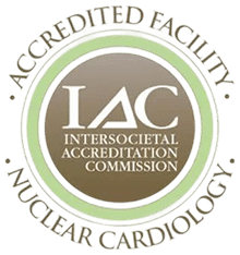
Міжтовариська акредитаційна комісія. Акредитований заклад. Ядерна кардіологія

Пов’язані місця

Знайдіть свого кваліфікованого фахівця

Ознайомтеся з нашою повною мережею лікарів, фахівців та медичних команд — використовуйте фільтри за послугами, мовою, місцем розташування та страховкою, щоб знайти те, що вам підходить.