
Imagen médica
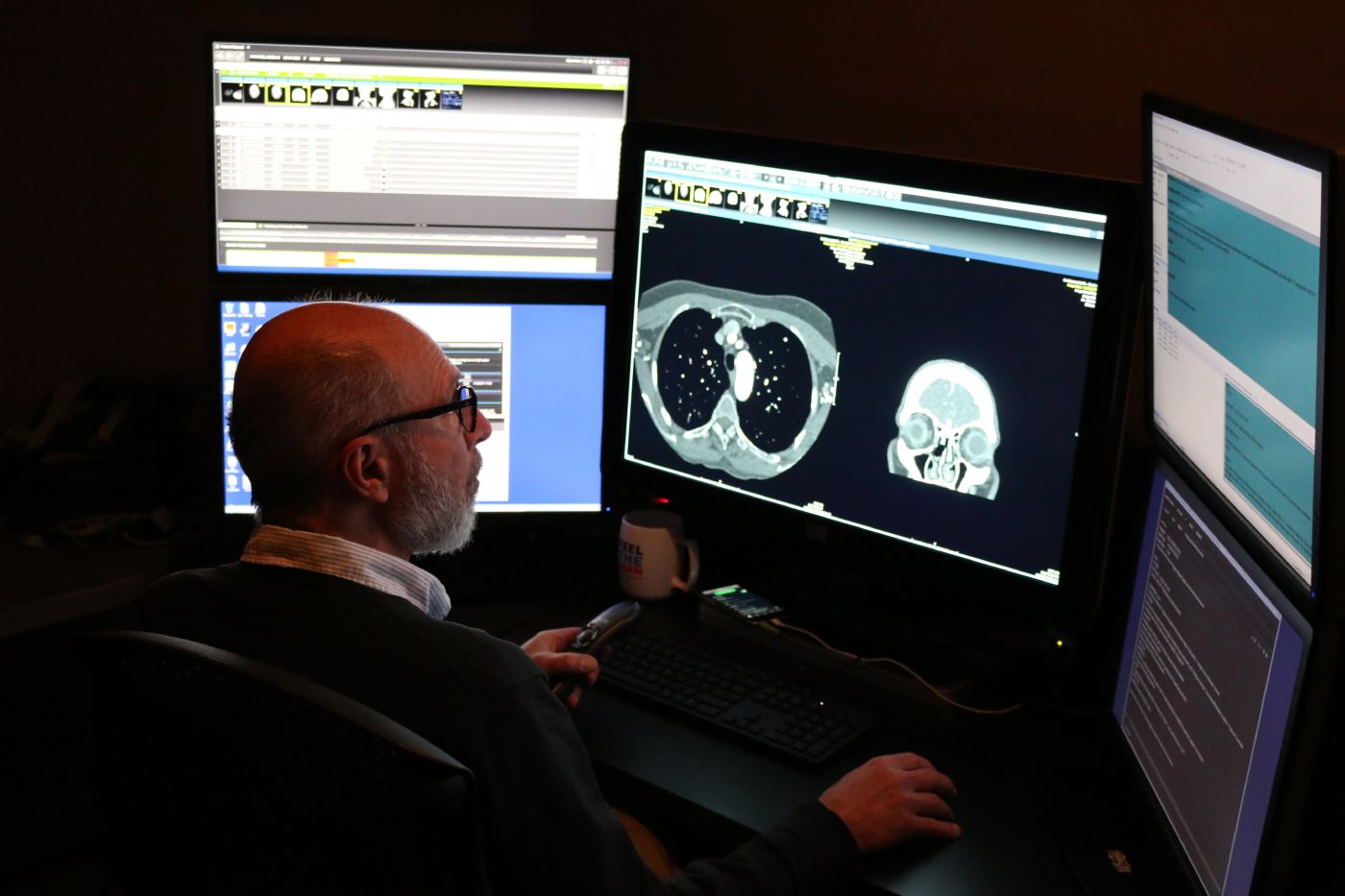
El servicio de diagnóstico por imagen utiliza tecnología avanzada para facilitar un diagnóstico preciso y la planificación del tratamiento. A veces, la clave para la curación reside en lo que no podemos ver a simple vista. Nuestro equipo de diagnóstico por imagen utiliza tecnología avanzada, como rayos X, resonancias magnéticas, tomografías computarizadas y ecografías, para ayudar a tus profesionales sanitarios a comprender lo que ocurre en el interior de tu cuerpo. Pero no se trata solo de máquinas, sino de personas que se preocupan por ti. Sabemos que las pruebas de diagnóstico por imagen pueden resultar intimidantes, por lo que nos tomamos el tiempo necesario para explicarle el proceso, responder a sus preguntas y garantizar su comodidad. Nuestros radiólogos y técnicos trabajan con precisión y empatía, ofreciendo resultados que orientan el diagnóstico y el tratamiento. Tanto si se trata de investigar un nuevo síntoma como de realizar un seguimiento de una enfermedad crónica, estamos aquí para ayudarle con claridad y amabilidad.
Podemos ayudarte con:
- Técnicas avanzadas de diagnóstico por imagen: tomografía computarizada (TC), resonancia magnética (RM) y tomografía por emisión de positrones combinada con tomografía computarizada (PET/TC)
- Diagnóstico por imagen mamario:mamografía, atención integral de la mama
- Salud ósea: DXA(prueba de densidad ósea)
- Imagenología especializada:Medicina nuclear
- Pruebas de imagen rutinarias:radiografía, ecografía
Frequently Asked Questions: CT Scan
A computed tomography (CT) scan is a noninvasive medical test that helps physicians diagnose and treat medical conditions. CT combines x-ray equipment with computers to produce multiple cross-sectional images of the body. These images can then be examined on a computer monitor.
The CT exam lasts about 10-15 minutes, unless you are drinking oral contrast or are having a specialized exam such as a biopsy or other procedure. Drinking oral contrast may add 30 minutes to two hours. Total time you will be at the healthcare facility is 30 minutes to 3 hours. This information will be available to you when you schedule your appointment. You should wear comfortable, loose-fitting clothing for your exam. You may be given a gown to wear during the procedure. Metal objects in the area being scanned, including jewelry, eyeglasses, dentures and hairpins, may affect the CT images and should be left at home or removed prior to your exam. You may also be asked to remove hearing aids and removable dental work. You may be asked to remove any piercings, if possible.
Es posible que se le pida que no coma ni beba nada durante unas horas antes de la prueba, dependiendo del tipo de examen. Es posible que se le coloque una vía intravenosa y que se utilice un medio de contraste intravenoso, o «tinte», durante la prueba. Debe informar a su médico de todos los medicamentos que está tomando y de si tiene alguna alergia. Si sabe que es alérgico al medio de contraste intravenoso, o «tinte», su médico puede recetarle medicamentos (normalmente un esteroide) para reducir el riesgo de una reacción alérgica. Por lo general, estos medicamentos deben tomarse 12 horas antes de la administración del medio de contraste intravenoso.
Inform your doctor of any recent illnesses or other medical conditions and whether you have a history of heart disease, asthma, diabetes, and kidney disease or thyroid problems. Any of these conditions may increase the risk of an unusual adverse effect to the contrast media. Women should always inform their physician and the CT technologist if there is any possibility that they may be pregnant.
En el caso de los pacientes pediátricos, se puede permitir la presencia de uno de los padres en la sala, pero deberá llevar un delantal de plomo o sentarse detrás de una barrera de plomo para minimizar la exposición a la radiación.
Frequently Asked Questions: Fluoroscopy
La fluoroscopia es una forma especial de radiografía. Mediante la fluoroscopia, las imágenes radiográficas pueden visualizarse como una secuencia de imágenes en movimiento, y el radiólogo puede observar órganos internos, como el colon, en movimiento. La fluoroscopia también ayuda al radiólogo a comprender cómo algunos órganos, como el estómago, vacían los alimentos que ingieres y a determinar si el estómago funciona correctamente. Los radiólogos y los técnicos en radiología cuentan con una formación específica para utilizar la dosis mínima de rayos X necesaria para obtener una imagen diagnóstica. Estos expertos saben cómo realizar pruebas de imagen tanto en adultos como en niños de forma segura.
Depending on the exam ordered, your physician may ask you not to eat or drink for several hours beforehand or provide specific preparation instructions. Some exams also require blood work to be completed before the imaging study. During your exam, you may be asked to change into a gown and remove jewelry, eyeglasses, dental appliances, or any metal objects or clothing that could interfere with the images. If there is any possibility you are pregnant, please inform your physician or technologist before the exam. Most imaging tests are avoided during pregnancy to limit radiation exposure. If an X‑ray is necessary, special precautions will be taken to protect you and your baby. Depending on the area being examined, images may be taken while you are standing or lying on an X‑ray table. Multiple images from different angles may be needed. Exam times typically range from 30 minutes to 2 hours.
For Barium Studies: If you do not have a bowel movement for more than 2 days after your exam, or you cannot pass gas through your rectum.
For Contrast Studies, complications are rare. If you were given contrast, call your doctor if any of the following occurs after the exam:
- Urticaria
- Picazón
- Náuseas
- Ojos o labios hinchados y con picor
- Opresión en la garganta
- Dificultad para respirar
When the examination is complete, you may be asked to dress and wait while the images are reviewed. Depending on the exam type, you may be asked to drink plenty of fluids to flush the barium or contrast out of your system. If you had barium, your stools may appear white, as your body clears the barium from your system. A radiologist will review the images and send a report to your referring doctor. You will receive your results from the doctor who ordered the test.
Frequently Asked Questions: X-Ray
Una radiografía es una prueba de diagnóstico por imagen que permite obtener imágenes del interior del cuerpo para ayudar a los médicos a diagnosticar afecciones médicas. Las radiografías más habituales incluyen imágenes del tórax, el abdomen, la columna vertebral, el cráneo, los senos paranasales y las extremidades. En algunas pruebas se utiliza un medio de contraste o bario para resaltar mejor determinados órganos o tejidos; estos estudios se realizan mediante fluoroscopia. Los radiólogos y los técnicos están especialmente formados para utilizar la menor cantidad de radiación necesaria para obtener imágenes nítidas y diagnósticas, así como para realizar pruebas de forma segura tanto en adultos como en niños.
Most x-rays require no special preparation. You may be asked to remove some or all of your clothes and to wear radiology attire during the exam. You may also be asked to remove jewelry, removable dental appliances, eyeglasses and any metal objects or clothing that might interfere with the x-ray images. Women should always inform their physician and x-ray technologist if there is any possibility that they are pregnant. Many imaging tests are not performed during pregnancy so as not to expose the fetus to radiation. If an x-ray is necessary, precautions will be taken to minimize radiation exposure to the baby.
When the examination is complete, you may be asked to dress and wait while the images are reviewed. A radiologist will review the images and send a report to your referring doctor. You will receive your results from the doctor who ordered the test.
Depending on the area of interest, you may be placed on the x-ray table, or the exam may be done standing. Various x-rays may be taken at different angles to best visualize the area. The duration of the exam ranges from 15 to 30 minutes, depending on the type of x-ray or number of images ordered by your doctor. Barium and contrast studies may take from 30 minutes to 2 hours.
Frequently Asked Questions: MRI
La resonancia magnética (RM) utiliza un potente campo magnético, ondas de radio y un ordenador para generar imágenes detalladas de las estructuras internas del cuerpo. Se utiliza para evaluar el cuerpo en relación con diversas afecciones, entre ellas tumores y enfermedades del hígado, el corazón y el intestino. La RM es una técnica no invasiva y no utiliza radiación ionizante.
MRI exams typically take 45 minutes to one hour per body part. If contrast is used, the exam may take an additional 15–20 minutes. Plan to spend one to two hours total at the facility. Little preparation is usually required. You may take your regular medications unless instructed otherwise. If fasting or other dietary restrictions are needed, you will be notified when scheduling. Please arrive 15–30 minutes before your scheduled exam time, as instructed during scheduling. You will change into radiology attire for your exam. Jewelry, hair accessories, and other metal items are not permitted. A locker will be provided for your belongings. If you have any implanted medical devices—such as electronic implants, vascular or cardiac stents, or other surgical implants—please bring your device card or related documentation. This information is required to confirm that the device is safe for MRI.
If you had IV contrast material, the technologist would remove the IV and place a bandage on the IV site. Drink plenty of fluids to help flush all contrast media out of your system. If you receive sedation, someone must drive you home. Your images will be read by a radiologist, and a report will be sent to your doctor. Your doctor will get results of your exam within a few days.
An MRI machine is a large, cylinder‑shaped magnet that is open at both ends. You will lie on a moveable table that slides into the center of the magnet for imaging. Before your exam, you will complete an MRI safety screening form. These questions may be asked when scheduling and will be reviewed again by the MRI technologist before your scan. If IV contrast is part of your exam, the technologist will place an IV beforehand. You will need to lie very still during the scan, as movement can blur the images and extend the exam time. Depending on the area being examined, you may be asked to hold your breath for up to 30 seconds at a time. The MRI suite is well lit and includes a fan for comfort. You can communicate with the technologist at any time using a two‑way intercom, and you’ll be given an alarm button to alert staff if you feel uncomfortable. During the exam, you will hear loud, intermittent knocking sounds. Earplugs or headphones will be provided, and you may choose to listen to music. If you experience claustrophobia, talk with your provider in advance. They may prescribe medication to help you relax, and you’ll need someone to drive you home if medication is used.
Las complicaciones son extremadamente raras (menos del 1 %).Si le han administrado un medio de contraste, llame a su médico si se presenta alguno de los siguientes síntomas después de la prueba:
- Urticaria
- Picazón
- Náuseas
- Ojos o labios hinchados y con picor
- Opresión en la garganta
- Dificultad para respirar
Frequently Asked Questions: Ultrasound
La ecografía es una prueba de diagnóstico por imagen que utiliza ondas sonoras para obtener imágenes del interior del cuerpo. Durante la prueba, se coloca sobre la piel un pequeño dispositivo manual llamado transductor y un gel. El transductor envía ondas sonoras al interior del cuerpo y los ecos que regresan se utilizan para generar imágenes en un ordenador. La ecografía no utiliza radiación. Dado que las imágenes se captan en tiempo real, las ecografías pueden mostrar el movimiento de los órganos internos, así como el flujo sanguíneo a través de los vasos sanguíneos.
Please wear comfortable, loose‑fitting clothing to your ultrasound exam. You may be asked to remove clothing and jewelry in the area being examined and change into a gown. Preparation varies depending on the type of ultrasound. Some exams require you to avoid eating or drinking for up to 12 hours beforehand. Others may require you to drink several glasses of water before your appointment and avoid urinating, so your bladder is full. You will receive specific instructions when your exam is scheduled.
During the exam, a trained ultrasound technologist (sonographer) will apply warm gel to your skin and move a small handheld device called a transducer over the area being examined. The transducer sends sound waves into the body and captures the returning echoes to create images. Multiple images may be taken by adjusting the transducer, and in some cases a radiologist may obtain additional images. Most ultrasound exams take 30 minutes to one hour to complete.
When the examination is complete, you may be asked to dress and wait while the ultrasound images are reviewed. After an ultrasound examination, you should be able to resume your normal activities immediately. A radiologist who specializes in ultrasound will review the images and send a report to your referring doctor. You will receive your results from the doctor who ordered the test.
Certificados y acreditaciones
Lorem ipsum dolor sit amet, consectetuer adipiscing elit, sed diam nonummy nibh euismod tincidunt.


Lugares relacionados

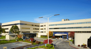
Hospital Confluence Health | Campus Central
1201 S. Miller St.
Wenatchee, WA 98801
Encuentra a tu proveedor especializado
Explora nuestra red completa de médicos, especialistas y equipos de atención; filtra por servicio, idioma, ubicación y seguro para encontrar la opción que mejor se adapte a tus necesidades.